BIO520 Exam 2 Spring 2010
Please email this lab to Jim Lund (jiml@uky.edu) with a subject line "BIO520 Exam 2" and name the document like so: "LundJ_exam2" or hand in written answers. Fill in your name on the exam!
You may use any books, notes, web pages, software programs, or related materials to complete this exam. You MAY NOT consult with any person regarding the exams intellectual content.
1. Examine the structure of human beta3 alcohol dehydrogenase, PDB ID 1HTB.
- a. (1 pt) What is the resolution of this structure?
- b. (1 pt) This protein structure was determined using X-ray diffraction. Would this protein be suitable for NMR structure studies? Briefly explain why or why not.
- c. (1 pt) Is this resolution sufficient to resolve only the protein backbone, individual residues, or atoms?
- d. (1 pt) How many protein chains are found in this structure?
- e. (1 pt) Does this structure file contain any atoms or molecules other than amino acids and water? Is so, briefly describe them.
- f. (2 pt) Does this structure file contain any atoms or molecules other than amino acids and water? Is so, briefly describe them.
- g. (2 pt) Which parameter measures how well the atomic model is supported by the experimental data? Also, what does the value of this parameter indicate about the quality of this structure (very poor, poor, worse than average, average, better than average, very good, exceptional)?
- h. (1 pt) Is this protein primarily alpha helix, beta sheet, or mixed?
- i. (1 pt) Along its longest dimension, what is the diameter of this complex? Include units in your answer.
- j. (2 pt) Which domains does this protein contain? Give the name, database, and ID for each domain you list.
3. (3 pts) Given a protein with homology only to other proteins of undetermined function, describe steps you could take to characterize it computationally. Give two things you attempt to predict about it and the program/analysis you would use.
4. (2 pts) Examine the energy plot of pairing for RNA sequence 'gcgtgcggctatgtgctagagggcaaagagcttcggttctatcttaggaaatgaagggcccgtggcacaagcgccgcaa' shown here: RNA dotplot. Base pair 1 is at the top left, bp 79 is at the bottom right. On the provided piece of paper 1) write your name and 2) draw this RNA structure. Ignore the sub-optimal lower energy matches (the colored sections).
5. The genomic sequence of a hydrogen sulfide producing bacterium from the human oral cavity, Veillonella dispar is sequenced independently by two companies, both to 10X coverage using shotgun paired-end whole genome sequencing. The completed genome is estimated to be 10 Mb. One uses Sanger sequencing and acquires reads with an average length of 900 bp and the second uses an Illumina sequencer and has sequence reads with an average length of 85 bp. As the total sequence is the same there are many more Illumina sequence reads.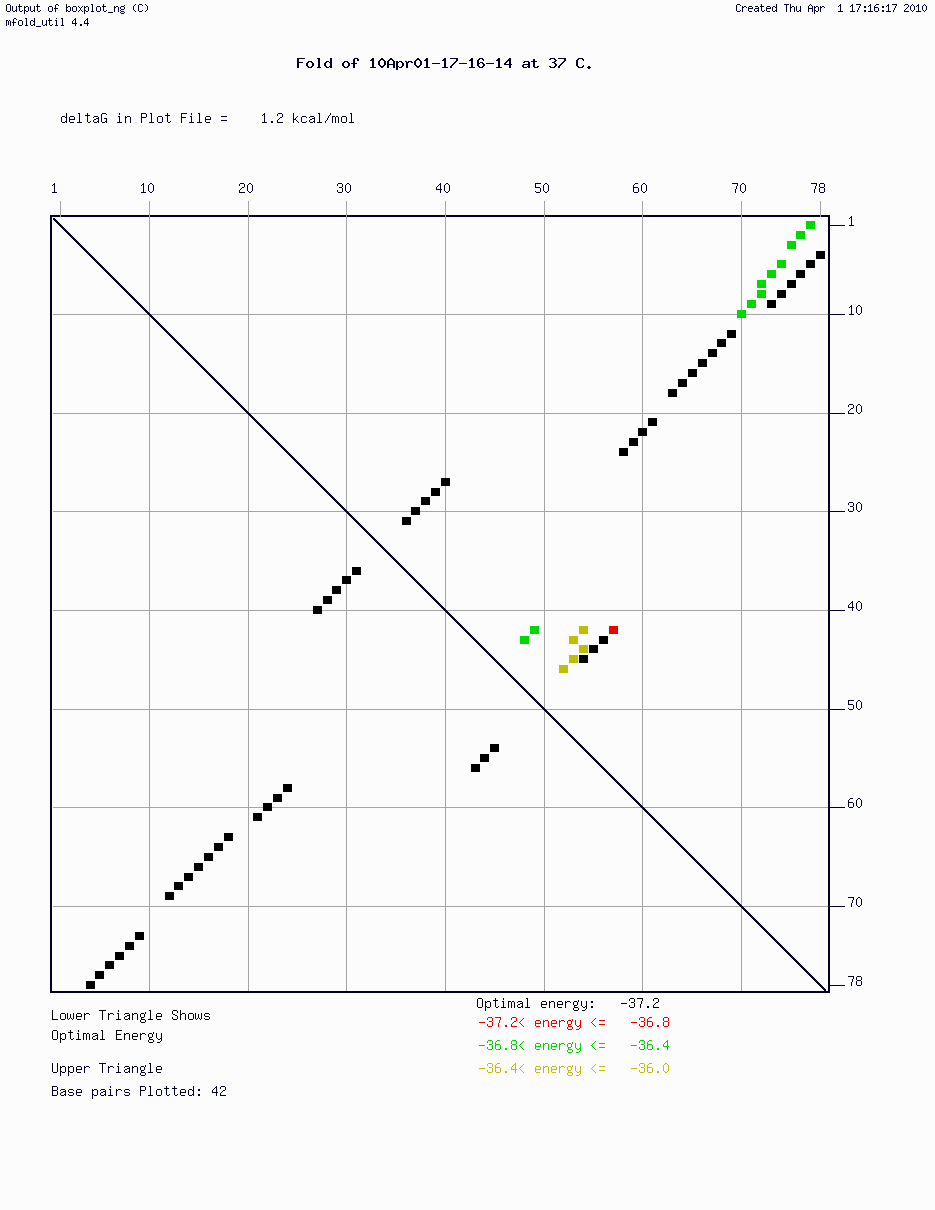
- b. (2 pt) Sequence assembly is done independently by both companies. How will the results of the sequence assembly from the Sanger data and the Illumina data differ?
- a. (2 pt) How many gaps do you expect from the Sanger sequencing vs. the Illumina sequencing?
6. (2 pt) The DOE sequencing finishing standards specify 99.9% per base accuracy. Briefly describe two serious problems that would arise from working with genomic sequence with a 99% accuracy, an average of one error in 100 bp.
7. Indicate which of the following is a part of gene annotation in each organism. Items can apply to one, both, or neither organism.
- a. (3 pt) gram-positive bacterium B. subtilus
- b. (3 pt) Giant panda
- a. PolyA site signal HMM.
- b. Databases of previously sequenced genes from the organism.
- c. BLAST searches of against other organisms.
- d. DNA secondary structure analysis.
- e. A splice donor hidden Markov model.
- f. ORF analysis.
- g. Codon bias.
- h. Depth of coverage.
- i. Shine-Delgarno element signal HMM.
- a. (3 pt) For each predicted gene, give the number of exons and the strand.
- b. (1 pt) Give the exon prediction that Genscan has the most confidence in. Either give the bps the exon covers or cut/paste the Genscan prediction line.
A. GeneMark
B. VAST
C. PDB
D. TRANSFAC
E. JPRED
F. TargetP
G. PSORT II
H. JMol
I. Cn3D
J. MFOLD
K. WoLF PSORT
L. Artemis
M. Phrap
N. Phyre
O. PDB
0. Predict membrane topology of proteins with membrane spanning α-helices.
1. Predict membrane topology of proteins with membrane spanning β-strands.
2. Protein threading server.
3. Program to assemble contigs from DNA sequences.
4. Uses homology modeling to predict protein structure.
5. Protein subcellular location.
6. DNA sequencing and assembly machine.
7. Protein structure database.
8. Phylogenetics trees--construction and analysis.
9. Transcription factor binding site database.
10. NCBI's protein structure viewer.
11. Web browser protein structure viewer
12. RNA folding.
13. Protein structure prediction web server.
14. RNA structure database.
15. De novo gene finding
16. Predict exon-intron gene structure in genomic DNA.
17. Predict protein secondary structure.
18. Subcellular localization prediction for eukaryotic proteins.
19. Subcellular localization prediction using signal sequences and homology.
20. Edit multiple sequence alignments.
21. Program for aligning protein structures.
22. View, edit, and analyze DNA annotations of genes and other sequence features.